Home
About
Services
Information
Results
Contact
Home
About
Services
Information
Results
Contact

Full Skin Check

Your appointment with Martin takes 15 minutes. First he'll ask you about your past medical history and risk of skin cancer, asking where you were born, about blistering sunburns, family history of skin cancer, sunbed and sunscreen use.

He will then examine your skin thoroughly from head-to-toe, including scalp, armpits and feet. He will ask about moles on your breasts, groin and buttocks and examine these areas if there are any concerns, with your consent. If you need a chaperone or would like a support person to be present, please let him know.

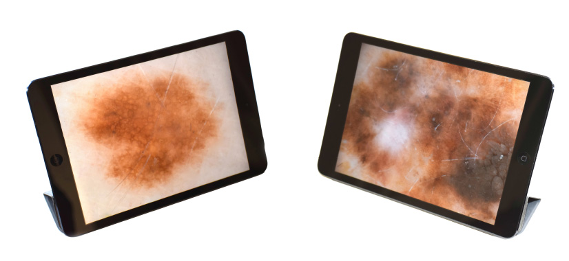
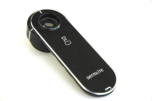
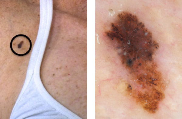
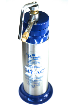
Martin performs your skin examination using a microscope called a dermoscope (shown below). Research shows that experts diagnose melanomas with only 60% accuracy with the naked eye. Using a dermoscope improves accuracy to over 90%. This is because the dermoscope enables the doctor to look beneath the top layer of the skin into the deep layer to reveal tell-tale signs of early melanoma and other skin cancers. Improved accuracy means less chance of missing a skin cancer and fewer benign lesions removed and sent to the lab unnecessarily, which saves money, pain, scars – and lives. Martin has completed years of dermoscopic training to interpret the special signs of early cancer.

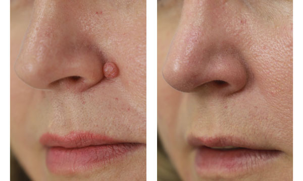
If Martin identifies any skin lesions of concern he will advise either a skin biopsy or complete surgical removal depending on the situation. This is usually booked about a week later and takes place at the same clinic under local anaesthetic. Martin performs all biopsies and surgical removals and is very gentle so the pain is no worse than a blood test.

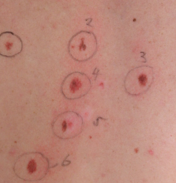
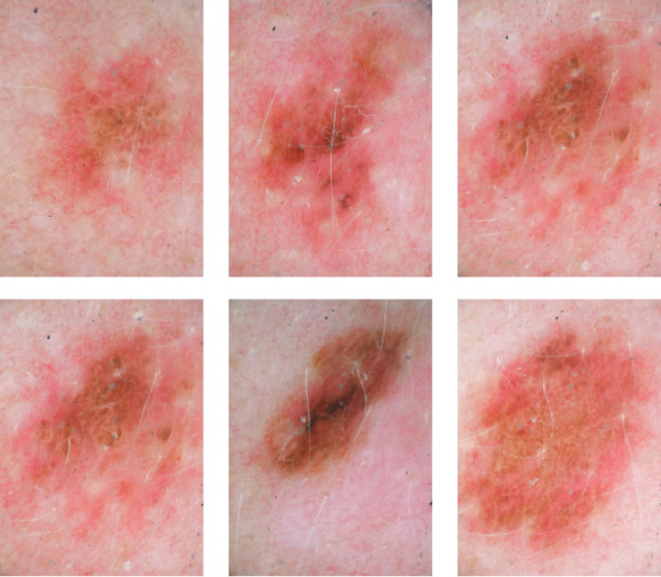
If you have a skin lesion that is not entirely normal and there is a low level of suspicion for melanoma, he will take a digital dermoscopic photograph of the lesion and advise you to come back three months later so he can rephotograph it and look for any changes. If there are no changes the lesion is over 99% likely to be benign. If there are changes he can surgically remove it.

Martin’s professionalism and thoroughness mean most people find skin checks to be an easy, reassuring experience.

Melanoma Screening

Skin examination involves looking with the naked eye and with a skin microscope called a dermoscope. Research shows that experts can only diagnose melanomas with 60% accuracy with the naked eye but with a dermoscope this increases to 90% or more. The dermoscope enables the user to look beneath the top layer of the skin (epidermis) into the deep layer (dermis) and can highlight tell tale signs of early melanoma and other skin cancer. Martin is a fully trained dermoscopist to Masters level and uses the latest 24-megapixel digital camera technology. The camera is wirelessly linked to an iPad so the images can be discussed with you straight away and a management plan decided upon before you leave the clinic. There is no waiting for reports.

Since early detection of melanoma is crucial, dermoscopic examination of the skin is essential. Martin uses digital photography to store images of your skin and highly magnified dermoscopic images of your moles. These images are used to compare your moles from year to year for early diagnosis of melanoma.

Here is an example of someone for whom digital dermoscopic melanoma screening is a good idea. This man has multiple abnormally shaped moles (atypical or dysplastic naevi) on his back. This increases his melanoma risk several times compared to the normal population. The photographs below show the incredible detail that the dermoscope can obtain. This is the gold standard for early melanoma detection.

Here is an example of a melanoma picked up using digital dermoscopy. The dermoscopic images are magnified to fill a 27- inch computer screen, which helps early melanoma detection.

Cosmetic Mole Removal

Cosmetic Mole Removal with Surgitron

Q. What is Surgitron®?

A. Surgitron® is a surgical electrode that uses high frequency radiowaves to provide incredible surgical control, precision, versatility and safety.

Q. What is Surgitron used for?

A. Surgitron® allows the removal of moles and other raised skin lesions on the face and body with virtually no risk of scarring. Removing moles on the face by surgical excision can leave a small, linear scar, no matter how experienced your surgeon. With Surgitron®, only the lesion itself is removed as surrounding tissues are not touched.

Q. What are the benefits of Surgitron®?

A. Virtually no risk of scarring because the removal is at a very superficial skin level. Minimal damage to surrounding skin due to high frequency radiowaves. No bleeding as radiosurgery cauterises as it cuts. Minimal, if any pain, due to the lack of heat damage to surrounding tissues. Rapid healing due to lack of tissue damage. Very low risk of post-operative infection as the electrode self-sterilizes.

Q. What should I expect when I have Surgitron?

A. A small amount of local anaesthetic is injected with a very fine needle under the mole. Immediately after removal, there is a raw, non-bleeding area, no bigger than the size of the mole. This forms a scab and 7-10 days later there is a fresh layer of new skin which blends with your skin colour over a few weeks.

Q. Which skin lesions can be treated?

A. Most raised skin lesions can be removed with Surgitron®; moles, skin tags, seborrhoeic keratoses, warts and verrucas.

Q. Which areas can be treated?

A. Face – there is minimal or no scarring, minimal or no pain and rapid healing. Facial veins - for people who are not suitable for laser, surgitron is a good alternative. Armpits, beneath breasts, neck – these are common sites for skin tags. Trunk, limbs – raised lesions anywhere on the body can be removed.

Q. What is the cost of treatment?

A. The cost of Surgitron® treatment depends upon the number of growths removed, but typically varies from NZ$400 to NZ$950.

Q. How do I make an appointment?

A. Please book an appointment with me at Silverdale Medical.

Liquid Nitrogen

Liquid nitrogen can be used to treat many benign, pre-malignant and malignant skin lesions. It is safe and effective if used according to guidelines. A fine spray applies it by using a cryotherapy gun like the one shown below. Liquid nitrogen is available at both clinics.

Liquid nitrogen is -195.8C and freezes the skin cells to -50C. This forms ice within the cells, causing cell death. The liquid nitrogen is sprayed for 3-30 seconds depending on the nature of the skin lesion and can be used to treat many lesions including sun damage, warts and some early or superficial skin cancers.

The treated area becomes red and slightly swollen, and then turns purple, brown then black. Clean the area daily with soap and water and don’t cover it. A scab forms, which you shouldn’t pick, but you can apply petroleum jelly. The scab usually falls off after 1-2 weeks on the face, 2-3 weeks on the hands and 4-12 weeks on the legs. It leaves you with slightly pink skin that then fades back to normal skin colour after a few weeks.

Complications with liquid nitrogen are very rare but include blistering, hypopigmentation (a white patch) and hyperpigmentation (a dark patch, more commonly seen in dark-skinned people).

Skin Cancer Creams

There are a number of creams available to treat selected pre-cancerous and cancerous skin lesions. Martin has extensive experience in using these treatments and can help you decide which is best for you.

Efudix Cream (5-fluorouracil)

Efudix cream has been used to treat sun-damaged skin since 1963. It was originally used as a chemotherapy agent and was found that a useful side effect was that it improved patients’ skin. It comes in a 20g aluminium tube with a 5-year shelf life. It is used to treat pre-cancerous skin lesions such as actinic keratoses and is applied 1-2 times daily for 3-4 weeks. For best results, different regions of the face can be treated at the same time e.g. forehead and temples. Do not treat an area greater than 23 x 23cm (the size of a dinner plate). Wash your hands after application and do not get it in your eyes, nose or mouth.

Treatment leads to redness, scaling, burning, itching and tenderness, which settles down once treatment stops. New skin grows over the treated area after 3-4 weeks. As with all of these topical treatments, there is a degree of “no pain, no gain”. Inflammation of the skin is needed to treat the sun damage so if the area is red and inflamed it means it’s working. As a guide, the inflammation takes 1-3 weeks to form, 3-6 weeks to settle and it takes 6-16 weeks for the sun-damaged skin to clear.

Aldara Cream (Imiquimod)

Aldara cream stimulates your immune system to treat pre-cancerous and some cancerous skin lesions. It comes in sachets, which should be used on the same day, but you can roll up the foil so they last a few days. Wash the area with soap, apply a thin layer of cream, rub it in, apply to a small margin of normal skin and leave on for 6-10 hours. It can be applied to an area 5x5cm = 25cm2. Avoid sunlight and tanning beds. Wear protective clothing and hats.

For actinic keratoses is applied 3 times per week (Monday, Wednesday, Friday) for 4 weeks. The area is assessed after another 4-8 weeks and if there are still lesions, the cycle can be repeated. For biopsy-confirmed superficial basal cell carcinoma it is applied 5 days per week (Monday-Friday with a break at the weekend) for 6 weeks. The area is assessed 6-12 weeks after treatment to see if it has been successful.

If there is too much redness or irritation, reduce the dose accordingly. You can use as infrequently as 2-3 days per week. The goal is to obtain tolerable amount of irritation, which is the evidence that your body is trying to fight the disease. If a severe local reaction occurs, such as extensive crusting or blistering, stop the Aldara cream until the reaction resolves. This usually takes 3-4 days, then restart at a reduced frequency.

Skin Biopsy

A skin biopsy is performed when there is concern that a skin lesion might be cancerous. A biopsy is the removal of a small piece of skin for examination under a microscope by a skin histopathologist. The procedure is done at both clinics. Local anaesthetic is carefully injected under the skin so the pain level is no worse than a blood test. A small piece of skin is then removed either with a punch biopsy tool or blade.

With a punch biopsy tool, a small 3-6mm circle of skin is removed and 1-2 stitches are put in to hold the wound together. The stitch is taken out a week or so later.

With a shave biopsy, a blade is used to shave a thin layer of skin. No stitches are needed.

A dry dressing is applied to the wound and you are asked to keep it dry for 24-48 hours. You can then have a shower with the dressing on. Remove the dressing after the shower and re-apply another dressing with some petroleum jelly to keep it moist (this speeds up healing). Both biopsies usually heal very well leaving minimal, if any scarring.

Skin Surgery

If there is a high index of suspicion for a melanoma, the mole is best removed entirely with a 2mm margin of normal skin. Basal cell and squamous cell cancers are removed with 3-5mm margins of normal skin. This is done at both clinics under local anaesthetic. The mole is removed as an ellipse as shown in diagram below. The length of the ellipse is usually 3-4 times the width. People often worry that the scar is too long for the size of the skin lesion but it helps the edges of the wound to oppose easily and enables a superior cosmetic result.

When a mole is removed from an area of skin tension (shoulders and back especially, but also trunk and limbs), deep sutures are placed to hold the wound together. These take 3-6 months to absorb depending on the type of suture material used. Sometimes surface sutures are also used. These need to be removed by a nurse 5-14 days after the procedure depending on the location of the surgery. Wound care is similar to skin biopsy. Keep it dry for 24-48 hours, and then you can run water over the wound. Don’t rub the wound with soap. Be gentle with it. Dab it dry and re-apply a dry dressing. Pain after the procedure is usually mild. Simple painkillers such as paracetamol are all that are usually needed. If there is any sign of redness, oozing or pus discharging from the wound, please contact me straight away as there could be a wound infection and a course of antibiotics might be needed. Martin’s risk of wound infection is very low at less than 1% of surgical cases.

Follow-up after skin surgery is essential so that the wound can be reviewed and the result of the histology (tissue examination) discussed. Histology not only confirms the diagnosis but also if the tumour has been completely removed. 97% of Martin’s malignant tumours are completely removed at the initial procedure.

If the mole removed is a melanoma, you will need to have a second procedure to take another margin of skin. This margin depends on the thickness of the melanoma and is usually 5-20mm. Patients often wonder why a doctor had ‘two attempts at it’ but this is standard practice because we need to make sure there is not a single melanoma cell left in your body. Re-excision is done at both clinics. In complex cases, Martin will refer patients to a Plastic Surgeon specializing in melanoma management.

Skin surgery will always leave a scar, no matter who performs the procedure. Scarring can be minimized with good surgical technique and post-operative wound care. Some people scar more than others and some areas of the body are more likely to scar e.g. upper anterior chest wall, shoulders and back. Most skin cancers are found on the head and neck and fortunately, the head and neck heals very well due to its excellent blood supply.

Corporate Services

Workplace skin checks

Martin regularly provides workplace dermoscopic skin cancer screening for companies. Your staff fill out a skin questionnaire and attend a 15-minute thorough head-to-toe skin examination. He takes the opportunity during the consultation to educate about the symptoms and signs of skin cancer, skin self-examination, sun avoidance and the use of sunscreens.

Skin cancer seminars

He also provides 30-40 minute interactive seminars on skin cancer aimed at teaching people about their risk profile, what to look for and how to reduce skin cancer risk. He has lots of interesting photographs and plenty of stories!

Pricing

Full Skin Check $250
Liquid Nitrogen $50
Skin Biopsy $300
Surgitron cosmetic mole removal $750
Skin Surgery $750-$1,500*

*Depending on the complexity of the operation.
We will provide you with a written quote once you have been assessed.

Southern Cross, NIB, Partner’s Life, Police, Veteran’s Affairs and other insurance companies affiliated.

BOOKING

Dr. Martin Denby • Skin Cancer Doctor